Monday, November 17, 2014

Week 4 Observations

This was the final week for lab observations and I was able to identify just a few more organisms one last time. This week especially, I noticed how much my micro aquarium has grown since we first started. My aquarium has expanded so much since the first week and in this process, I've tried to identify as many organisms as I could. My aquarium definitely has become more familiar which all the previous organism's I have identified I have started to repeatedly see them throughout.  
Image 1. This specific species of seed shrimp has been identified as a Darwinula which is further described in "Guide to Micro Life" by Kenneth G. Rainis on pg. 211 image 6. This was the only seed shrimp that I saw and it did not really move when I was observing it. 

Image 2. This organism has been identified as Cladophora which is further described in "The Freshwater Algae" by G.W. Prescott on pg. 132 Fig. 218. This organism was found by Professor McFarland in my micro aquarium and this was also another organism that did not have noticeable movement so initially I was not even sure if it was an organism or not.
Image 3. This organism is identified as Vorticella in "Free-Living Freshwater Protozoa: A Color Guide" by D.J Patterson on pg. 113 Figure 232. I have seen quite a few Vorticella throughout my micro aquarium as they are easy to observe since there is not really movement among them.


Bibliography

Prescott, G.W. The Fresh-Water Algae. W.M.C Brown Company Publishers.

Rainis, Kenneth G., Russel, J. Bruce. Guide to Micro Life. Danbry, CT. A Division of Groller Publishing.

Monday, November 10, 2014

Week 3 Observations

Even after the third week, I am continuing to identify new organisms with the help of Professor McFarland. Im starting to see how diversitized my micro aquarium really is as there seem to be new organisms every week. I also keep seeing previous organisms that I have already identified but I continue to watch how they interact in the micro aquarium every week to see if they change their habits. 

Image 1. This organism is a Peranema which is further described in  Free-living Freshwater Protozoa: A Color Guide by D.J Patterson on pg. 51, figure 70. I did not see many of these organisms but I was able to easily spot it out as the Peranema moved at a steady pace so I was able to observe it. 

Image 2. This organism has been identified as an Epalxis which is further described in Free-living Freshwater Protozoa: A Color Guide by D.J Patterson on pg. 141, figure 311. This organism was hard to get a picture of (hence the blurry picture) as it moved around at a very abrupt and fast pace. Luckily I was able to get a picture, because if not I may not of had another chance in capturing the Epalxis.


Bibliography
(Both sources have already been cited in previous blogs)


Monday, November 3, 2014

Observations for Week 2

Week 2: This week Professor McFarland put a a fish food pellet from Atison's Betta Food (information below) into each micro aquarium and with this, I observed a lot of organisms around this food pellet and also was able to identify a few more new organisms. My micro aquarium is starting to become more familiar as I see a lot of the organisms that I identified last week all around the micro aquarium plus the ones I just identified. There were also a few more organisms that I found this week that I was unable to identify because they were either moving around the micro aquarium too fast or were hidden so I could not see enough  of the organism to be able to identify what it is. With this, I was still able to observe new things and the images below are what I found for this week.

"Atison's Betta Food" made by Ocean Nutrition, Aqua Pet Americas, 3528 West 500 South, Salt Lake City, UT 84104. Ingredients: Fish meal, wheat flower, soy meal, krill meal, minerals, vitamins and preservatives. Analysis: Crude Protein 36%; Crude fat 4.5%; Crude Fiber 3.5%; Moisture 8% and Ash 15%.

Image 1. Identified organism is a Nematoda as found in Fresh-Water Invertebrates of The United States: Protozoa to Mollusca by Robert W. Pennak pg. 228 fig. 1. In this picture I was only able to get the posterior end of the Nemoda as it was swimming around very rapid and then its anterior end was hidden behind a leave for a while so I could only observe this aspect of the Nematoda.

Image 2. The Craspedacusta Sowerbyi was also identified in Fresh-water Invertebrates of the United States: Protozoa to Mollusca by Robert W. Pennak pg. 116 fig 5.b. This was the only one I saw of this organism as professor McFarland told me it is a freshwater Jellyfish. There was no movement along with it as it is in one of its earlier developing stages so I am assuming that is why there was not a great amount of movement. The diagram in the book showed the stages of the Craspedacusta Sowerbyi's growth cycle and identified this organism in one of its earliest stages so hopefully I will be able to continue to watch it develop. 

Bibliography:

Pennak, Robert W. Fresh-Water  Invertebrates of the United States: Protozoa to Mollusca. 3rd ed. A Wiley-interscience publication. 

McFarland, Kenneth [Internet] Botany 111 Fall 2014. [15 October 2014]. Available fromhttp://botany1112014.blogspot.com/


Sunday, October 26, 2014

Observations for week 1

Week 1: I was able to observe many new organisms that I did not see my first week when I initially set up my micro aquarium. There were multiple protozoa and  a diatom that I was able to identify due to Professor McFarland's help. I spent a good amount of time trying to identify and watch each organism under the microscope and understand how each one moved about and participated within the micro aquarium. There were a few organisms that I observed but have yet to be identified as they were moving too quickly within the micro aquarium for me to be able to take a picture of them. But from the ones I did observe and identify each organism had very unique traits from one another including how they moved about in the aquarium, which is further described under each figure. 


 Image 1.  Identified protozoa is Anisomena sp. as shown in Free-living Freshwater Protozoa: A Color Guide by D.J Pattersonpg. 54, Figure 78. Anisomena are abundant throughout my micro aquarium as I observed  multiple of them. They are not known to have any ingestion so with that, they absorbed wastes that they cannot rid of which I watched them do under the microscope as I saw this protozoa take in substances that you could still see within it.

 Image 2. Identified protozoa is Euplotes sp. as shown in Free-living Freshwater Protozoa: A Color Guide by D.J Patterson, pg. 124, Figure 260. Also saw multiple of these organisms throughout my micro aquarium and learned that there use their central cirri for movement. The central cirri I learned in Patterson's book are the little white legs that you can see in image 2 and when I observed this organism I was the most intrigued by their legs and how they moved around with as it was quite the sight to see.

 Image 3. Identified diatom is Nitzschia Paradoxa as shown in Diatoms of North America by William C. Vinyard on pg. 178 ,Figure 141. I observed the Nitzschia Paradoxa for a while as this was the only one I saw and I waited to watch as its parts dissembled and the Nitzchia Paradoxa went from being a mass to a thin line as it pulled apart. The Nitzschia Paradoxa at first did not move a lot until it started to change shapes and dissemble and that is when I saw a lot of spastic movement from this diatom.


Bibliography

Patterson, D.J. 2013. Free-Living Freshwater Protozoa: A Colour Guide. 5th impression. London (UK): Manson Publishing Ltd.

Vinyard, William C. Diatoms of North America. Eureka, CA: Mad River Press Inc. 

Sunday, October 19, 2014

Observation 1

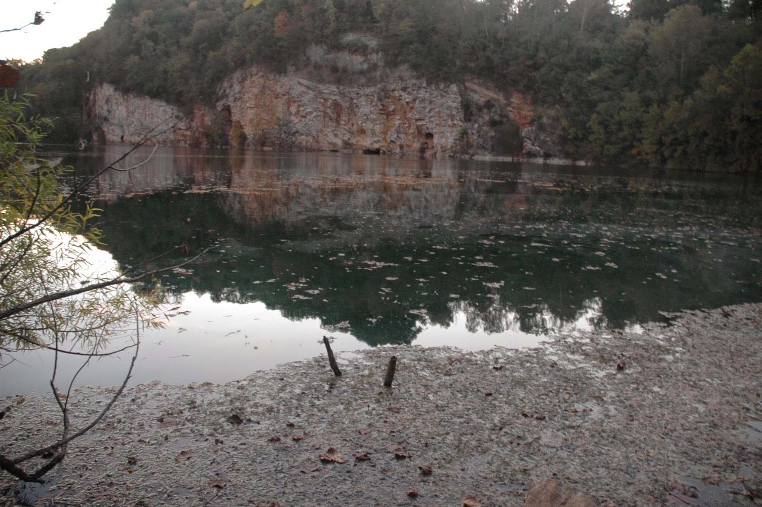
Water Source used: #5 Meads Quarry, Island Home Ave

5. Meads Quarry, Island Home Ave
5. Meads Quarry, Island Home Ave, Knox Co. Tennessee Partial shade exposure Rock Quarry N35 57.162 W83 51.960 880 10/12/2014


Lab section:blue
Table: red
seat number: blue

Procedure:

On 10/15/14 the process of setting up and starting the micro aquarium experiment began by first setting up the micro aquarium itself. This was done by adding a base to hold up the aquarian and then a lid to seal the water in. After this, each student was given a 3 sticker color combination to clarify each aquarian from another. My micro aquarium specifically had a blue, red, and another blue sticker on the left side and each sticker was labeled with my initials (PA) to make it more distinct from the other lab students aquariums. After this, each student chose their own water sample and applied a sample into the aquarium. My water example came from water source 5 otherwise known as the Meads Quarry. In order to put a diverse water sample into the aquarium, water was extracted from the dirt and mud at the bottom of the Meads Quarry water source, off of the leaf extracts in the water and then just off of the surface of the water. All these samples were dripped into the micro aquarium. After that, two water weeds were added to the micro aquarium to oxygenate the water. After all these aspects from the water source were added to micro aquarium we then observed our samples under a microscope to begin to understand was is in our micro aquariums.

Observations: 
When observing my micro aquarium, there seemed to be multiple unidentified one celled organisms swimming around. They were a dark brown color and would move around very quickly and seemed to be swimming around the water mosses and were hidden behind the mosses at certain points. While observing their actions, these organisms seemed like they were eating/collecting substances from the water as it looked like they would vacuum these substances into them. 

Information on the plants used in the Micro Aquarium:

Amblestegium varium (Hedwig) Lindberg. Moss.
Collection from: Natural spring. at Carters Mill Park, Carter Mill Road, Knox Co. TN. Partial shade exposure. N36 01.168 W83 42.832. 10/12/2014


Utricularia gibba L. Flowering plant. A
carnivorous plant. Original material from south shore of Spain Lake (N 35o55 12.35" W088o20' 47.00), Camp Bella Air Rd. East of Sparta Tn. in White Co. and grown in water tanks outside of greenhouse at Hesler
Biology Building. The University of Tennessee. Knox Co. Knoxville TN.
10/12/2014